
Advancing Cardiac Care
Consistency: The Hidden Challenge in Echocardiography
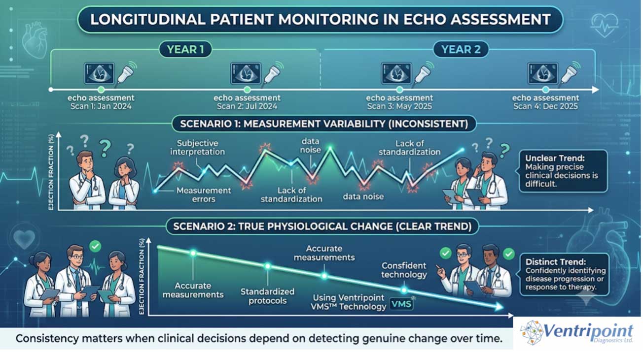
Echocardiography is the most widely used imaging modality for assessing ventricular function and plays a vital role in routine patient management. Increasingly, clinical decisions are based not on a single scan, but on measurements collected over months or years as part of ongoing patient monitoring.
For these measurements to be meaningful, clinicians need confidence that changes in ventricular volumes or function reflect true physiological change rather than differences in how the study was acquired or analysed. Variability can arise at multiple stages of the workflow, including image acquisition, landmark identification, and measurement methodology.
As healthcare becomes increasingly data-driven, reproducibility is becoming just as important as accuracy.
For patients undergoing long-term follow-up, consistent measurements provide a stronger foundation for identifying trends, guiding treatment decisions, and delivering the best possible care.

The PLUS of VMS+
AI-Assisted Landmark Placement: Supporting Consistency in Cardiac Imaging
Measurement variability in echocardiography can arise at multiple stages of the workflow, from image acquisition through to landmark identification and border tracing during analysis.
When assessing ventricular volumes and ejection fraction, small differences in how cardiac structures are identified can influence the final measurements generated from a study. For serial patient follow-up, reducing avoidable sources of variability becomes increasingly important.
One area where standardisation can add value is anatomical landmark placement.
In traditional workflows, clinicians identify anatomical landmarks manually as part of the analysis process. While these measurements remain clinically useful, minor differences in landmark placement can contribute to variability between studies.
The VMS+ 4.0 AI-assisted workflow was developed to support greater consistency by automatically identifying and suggesting anatomical landmarks during analysis. Rather than replacing clinician expertise, the software provides a standardised starting point that can be reviewed, verified, and adjusted by the user as needed.
The Ventripoint whitepaper, “Use
of AI to Aid 2D Echo Interpretation: VMS+ 4.0 vs. 3.0,” demonstrated how AI-assisted point placement can streamline analysis workflows while improving efficiency and supporting reliable measurement generation.
The enhanced workflow reduces the amount of manual interaction required during analysis while retaining full clinical oversight.
Key benefits of the AI-assisted workflow include:
- Standardised landmark identification
- Reduced manual workflow burden
- Improved analysis efficiency
- Consistent application of analysis methodology
- Greater confidence in longitudinal patient assessment
As AI adoption continues to expand across cardiovascular imaging, its greatest impact may not simply be automation—but helping clinicians achieve more reproducible and consistent assessments over
time.
Tech+
Three Practical Ways to Improve Consistency in Serial RV Assessment
While technology can support reproducibility, consistency begins with good clinical practice.
1. Standardise Image Acquisition
Use consistent imaging views, acquisition protocols, and patient positioning whenever possible. Variability introduced during image acquisition can influence all downstream measurements.
2. Apply Consistent Analysis Methodology
Using the same analytical approach across studies improves comparability and reduces variation that may arise from differences in measurement technique.
3. Focus on Reproducible Landmark Selection
When analysing serial studies, consistency in identifying key anatomical landmarks can help improve confidence that observed measurement changes reflect true physiological differences.
Small improvements in consistency at each step of the imaging pathway can have a significant impact on long-term patient monitoring.

Case Study / Clinical Perspective
Why Reproducibility Matters More Than Ever
Imagine a patient with repaired Tetralogy of Fallot returning for annual follow-up.
The clinical question is not simply:
“What is the right ventricular
volume today?”
The more important question is:
“How has the right ventricle
changed since the last study?”
When serial imaging is used to guide intervention timing and monitor disease progression, clinicians depend on measurements that are not only accurate, but comparable over time.
This is why reproducibility has become a growing focus across cardiac imaging. Reliable longitudinal assessment allows clinical teams to make decisions with greater confidence and helps ensure that patient management is guided by true physiological change rather than measurement variation.
Looking Ahead
As cardiac imaging continues to evolve, the conversation is increasingly moving beyond the question of accuracy alone.
The future of echocardiography will depend on delivering measurements that are not only accurate, but also reproducible, efficient, and meaningful across the entire patient journey.
By supporting standardised workflows and consistent analysis, advanced imaging tools can help clinicians focus on what matters most: identifying true change and delivering the best possible patient
care.
Community Spotlight
Supporting CHD Teams Beyond the Scan
Our clinical and research partners continue to shape how VMS+ evolves — from validation studies to discussions around scalability and workflow integration. Your insight helps support sustainable, high-quality cardiac imaging.
Join the VMS+ Movement
Interested in seeing how intuitive 3D reconstruction can fit into your echo workflow?
- Request a Demo— explore VMS+ in your own
clinical environment - Share Your Experience — your insights
shape our roadmap - Connect With Us— let’s advance cardiac
care together
Let’s transform cardiac care together: one patient, one insight, one workflow at a time.
Warm regards,
Matt Dobson,
Marketing Director
Ventripoint Diagnostics Ltd
VMS+ is for trained Healthcare Professional use only.
Availability subject to regulatory approval in respective regions: please contact Ventripoint Diagnostics Ltd. for further information.