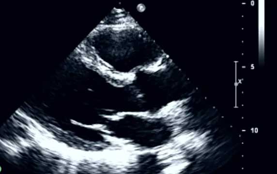
VMS+

VMS+: Revolutionizing Cardiac Care

We proudly introduce the VMS+, our groundbreaking step in cardiac imaging that transforms 2D echocardiograms into precise 3D heart models. This tool not only sets new standards in diagnostic accuracy but also fits seamlessly into existing clinical workflows, redefining cardiac care. Join us in exploring the capabilities and benefits of the VMS+ system on our website.

Fast, accurate and reproducible measurements

Harnessing the power of KBR, VMS+ is designed to create an accurate 3D model of the heart from a 2D echocardiogram and provides cardiac metrics on all four chambers of the heart within minutes. Obtain accurate results that do not require “perfect images”

View a 3D model of the heart.

Our proprietary Knowledge Based Reconstruction (KBR) algorithm leverages a MRI shape catalogue and anatomical landmarks from standard 2D echocardiogram images to construct a complete 3D model of the heart.

Security is our priority

Ventripoint’s system is highly secure. All diagnostic data created stays within an institution’s own IT and server system by default

Insights into the heart's structure and function for all conditions

Innovative sensor tracking system

This small, innovative device attaches to the ultrasound probe via a small adaptor, enabling patient repositioning during the examination without affecting results

Remote Installation

The VMS+ is designed to be immediately operational and integrated in the hospital’s infrastructure.

Results in 3 easy steps

Integrating the VMS+ into your workflow is quick and easy. Analysis takes minutes. The system simply needs images collected in a typical 2D echo exam and is compatible with any commercially available 2D ultrasound system.

1. Acquire Images

Accepts the ultrasound images from a video output of the 2D ultrasound and tracks the ultrasound transducer’s 3D spatial coordinates and orientation via our sensors during echo scan.

2. Place points

Points are placed on the images to mark the position of anatomic landmarks. No tracing is required. Takes just 5 minutes to complete.

3. Generate Results

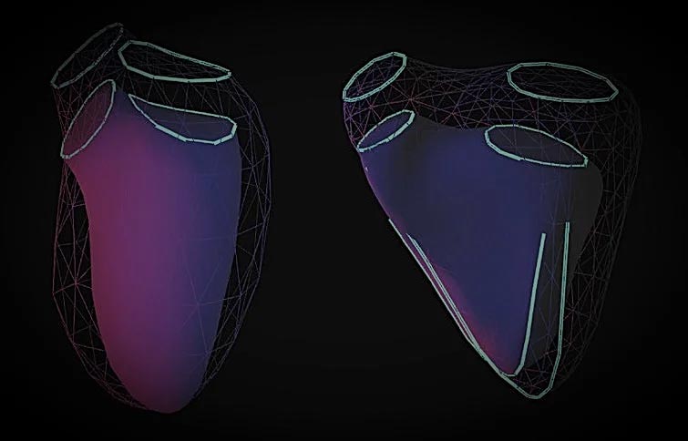
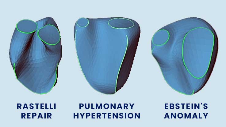
Volumes and ejection fractions are then calculated for the ED and ES phases of the cardiac cycle and the 3D heart can be viewed on different planes. All study files can be stored on PACS.

Want to talk to us about VMS+?

Talk to a representative to learn how VMS+ can be used to aid in more accurate volumes and ejection fractions so you can follow patients with confidence.

The Echo A.I. Alternative to cMRI