
Depth of heart, depth of care May 2026
Advancing Cardiac Care
As demands on echocardiography labs continue to grow, the need for tools that are not only accurate, but genuinely easy to use, has never been more important. This month, we focus on usability — how thoughtful design enables advanced cardiac imaging to become a seamless part of everyday clinical care.
The PLUS of VMS+
Designed for Clinicians: What the VMS+ Usability Study Tells Us
Advanced imaging only delivers value when it fits naturally into clinical workflows. To ensure this, Ventripoint conducted a formal human factors and usability study of VMS+ 3.0, evaluating how real clinicians interact with the system in realistic echo environments.
The study was designed to assess effectiveness, efficiency, and user satisfaction, validating that VMS+ supports safe, intuitive, and confident use — even for first-time users.
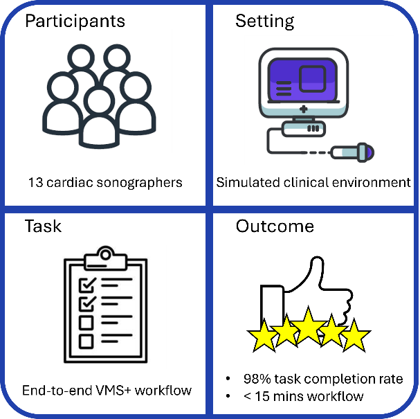
Study Overview
- 13 cardiac sonographers and healthcare professionals
- Representative of typical VMS+ users, with broad echocardiography experience
- No prior hands-on experience with VMS+
- Simulated hospital echo environment
- Standard clinical tasks: acquisition, analysis, 3D visualization, and reporting
Participants were trained using the same approach provided to new customers, followed by a short delay to reflect real-world training retention.
 Key Results
- 98% task completion with no critical errors
- Average workflow completed within 15 minutes
- High ease-of-use and confidence ratings after just one training session
Clinicians consistently highlighted how VMS+ supports accurate ventricular volume and ejection fraction measurements — particularly for the right ventricle — without disrupting standard echo acquisition routines.
“Makes you a better sonographer because you think about the landmarks when scanning.” — Usability study participant
Why This Matters
The usability study confirms what clinicians need most from advanced echo tools:
- Minimal clicks and data entry
- Clear guidance during acquisition and analysis
- Easy integration alongside the ultrasound machine — not between clinician and patient
- Rapid access to reproducible, quantitative results
By aligning directly with existing workflows, VMS+ enables teams to access cMRI-comparable volumetric insights without the operational barriers of MRI scheduling, sedation, or extended scan times.
Tech+
From Scan to Insight: The VMS+ Three-Step Workflow
Advanced technology delivers clinical value when it is clear, repeatable, and easy to remember. At the core of VMS+ usability is a simple three-step process that mirrors how clinicians already perform echocardiography — reducing cognitive load while enabling advanced 3D quantification.
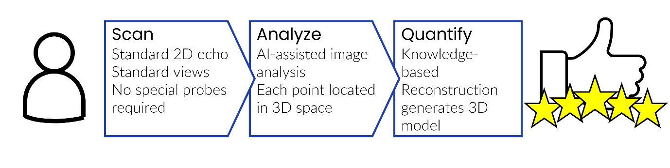
Step 1: Acquire
VMS+ begins with standard 2D echocardiographic acquisition using familiar views. There is no need for complex new scanning techniques or changes to established protocols. The system tracks probe position automatically, allowing clinicians to focus on image quality and patient interaction.
Why it matters: Consistent acquisition reduces variability at the source and ensures high-quality data for reconstruction.
Step 2: Analyze
In VMS+ 4.0, analysis is accelerated with AI-assisted point placement. Rather than asking clinicians to manually trace or contour the endocardial borders — a time-consuming and highly operator-dependent task — VMS+ uses a small number of key anatomical landmarks to define cardiac geometry. The system automatically identifies and proposes these landmarks based on learned patterns from validated datasets, while clinicians remain fully in control to review, adjust, and confirm.
Why it matters: Landmarking reduces the need for repetitive border tracing, lowering cognitive load and inter-observer variability. By anchoring analysis on consistent anatomical reference points, AI assistance shortens analysis time while supporting accurate, reproducible quantification — even in complex ventricular anatomy.
Step 3: Quantify
Using Knowledge-Based Reconstruction, VMS+ generates a 3D model of the heart, delivering volumetric measurements and ejection fractions derived from MRI-validated shape libraries, for all four chambers: RV, LV, RA and LA.
Why it matters: Clinicians receive rapid, quantitative results that support confident longitudinal assessment — without the access and scheduling barriers of cMRI.
Designed for Real-World Echo
The VMS+ three-step process was validated in the usability study, with nearly all participants completing the full workflow efficiently after a single training session. By aligning advanced reconstruction with intuitive steps — acquire, analyze, quantify — VMS+ turns sophisticated 3D analysis into a natural extension of everyday echo practice.
Community Spotlight
Designed With Clinicians, Refined by Experience
Insights from this usability study — together with continuous feedback from our clinical partners — directly shape how VMS+ evolves. From UI enhancements to guided workflows, clinician input remains central to every release.
We’re grateful to the sonographers and cardiology teams who help ensure that innovation translates into meaningful improvements at the bedside.
Join the VMS+ Movement
Interested in seeing how intuitive 3D reconstruction can fit into your echo workflow?
- Request a Demo — explore VMS+ in your own clinical environment
- Share Your Experience — your insights shape our roadmap
- Connect With Us — let’s advance cardiac care together
Let’s transform cardiac care together:
one patient, one insight, one workflow at a time.
Warm regards,
Matt Dobson,
Marketing Director
Ventripoint Diagnostics Ltd
VMS+ is for trained Healthcare Professional use only.
Availability subject to regulatory approval in respective regions: please contact Ventripoint Diagnostics Ltd. for further information.

